3.2.9 Triplanar
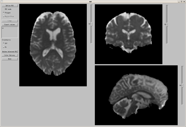
The triplanar viewer allows the user to view axial, sagittal and coronal views simultaneously. It also provides ROI drawing tools which can be used on any of the image orientations.
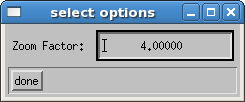
First, to set the magnification factor, use the opt button beside triplanar. The default value is 4, and is generally acceptable for most human brain data:

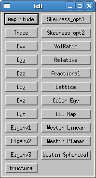
Two windows will appear when you click the triplanar button, a viewer and an image type menu.


Image Type Menu
Image Types
These images are explained in detail in the section describing the axial-only ROI utilities.
- Amplitude: a calculated reference (S0) image, calculated from the intercept of the tensor fitting.
- Trace: the trace of the diffusion tensor. Mean diffusivity (MD) = 1/3(TRACE)
- Dxx, Dyy, Dzz: diagonal tensor elements
- Dxy, Dxz, Dyz: off-diagonal tensor elements
- Eigenv1, Eigenv2, Eigenv3: the eigenvalues
- Structural: The image used as target for registration (if it exists)
- Skewness_opt1:
- Skewness_opt2:
- VolRatio: volume ratio
- Relative: relative anisotropy
- Fractional: fractional anisotropy
- Lattice: lattice index
- Color_Egv: a color coded eigenvalue image
- DEC Map: directionally encoded color maps
- Westin Linear: Westin linear measure
- Westin Planar: Westin planar measure
- Westin Spherical: Westin spherical measure
The menu on the left hand side of the viewer provides display and ROI drawing options. ROI options are discussed in the next section. Display options are noted with red boxes below.


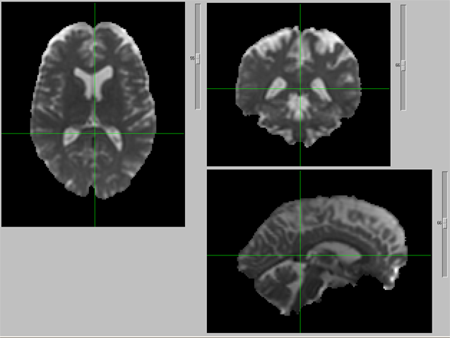
- Crosshairs: when turned on, the cross hairs will be placed where the mouse cursor is clicked in the image. It is placed on all 3 orientations, and is useful for identification of anatomical structures in all 3 orientation views.
- The color options are identical to the color options for the roi utilities.
- Use the Done button to exit the triplanar viewer.
